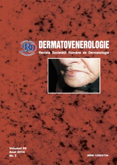
Carcinomul scuamos (CS) de buza este o tumora maligna de origine epiteliala, infiltranta si distructiva, cu potential de metastazare pe cale limfatica si/sau sanguina.
El reprezinta 15-30% din CS ale extremitatii cefalice si 1/5 din cancerele tractului aerodigestiv superior.
Noi am efectuat un studiu clinic, histopatologic si imunohistochimic pe un lot format din 43 bolnavi spitalizati si tratati in Clinica Dermatologie Craiova in anii 2013-2014.
Bolnavi si metoda. Fiecarui bolnav i s-a intocmit o fisa, cuprinzând date de identificare, mediul de provenienta, profesia, factorii de risc, istoricul bolii, localizarea si diametrul tumorii, forma clinica, rezultatul examenului histopatologic si al investigatiilor imunohistochimice.
Pentru studiul histopatologic piesele au fost prelucrate prin tehnica clasica de includere la parafina si colorate cu hematoxilina-eozina. Evaluarea histopatologica a vizat: varietatea carcinomului scuamos; gradul de diferentiere; prezenta invaziei perineurale si perivasculare; asocierea cu leziuni displazice la periferia tumorii; prezenta celulelor maligne reziduale la nivelul marginilor de siguranta chirurgicala.
Am efectuat investigatii imunohistochimice la zece cazuri, utilizând ca anticorpi p53, VEGF si Ki67. Am folosit sistemul de vizualizare LSAB2 (Dako, cod K0675).
Developarea s-a realizat cu ajutorul cromogenului 3,3’- diaminobenzidina tetrahidroclorid (Dako, cod K3468).
Controlul negativ s-a realizat prin omiterea anticorpului primar.
Rezultate
In toate cele 43 de cazuri, tumora a fost localizata la nivelul buzei inferioare. Dintre acestea, 31 de cazuri le-am intâlnit la barbati, restul celor 12 cazuri au fost prezente la femei. Din rural proveneau 37 de cazuri. Vârsta bolnavilor a fost cuprinsa intre 40 si 87 ani (vârsta medie 70,86 ani).
Forme clinice: CS vegetant ulcerat (13 cazuri), ulceroinfiltrativ (10 cazuri), nodular (9 cazuri), keratozic (5 cazuri), ulcerat (4 cazuri), carcinom in situ (2 cazuri).
Sub aspect histopatologic a predominat CS moderat diferentiat (23 cazuri), urmat de varianta bine diferentiata (13 cazuri) si slab diferentiata (2 cazuri). La 3 bolnavi am intâlnit carcinom verucos si ceilalti doi bolnavi prezentau carcinom in situ. Invazia perineurala si perivasculara a fost prezenta la 4 bolnavi, iar la 26 de cazuri au fost prezente modificari de tip displazic la periferia tumorii. Am intâlnit la 5 bolnavi celulele maligne reziduale la nivelul marginilor de siguranta chirurgicala.
Investigatiile imuno-histochimice au relevat ca marcajul pozitiv VEGF a fost prezent in toate cazurile studiate, expresia fiind intens pozitiva la 8 cazuri (80%), moderat pozitiva la 2 cazuri (20%). Marcajul p53 este nuclear si a fost observat in toate cazurile, intens pozitiv difuz in 6 cazuri (60%) si pozitiv focal in 4 cazuri (40%). Marcajul Ki-67 este nuclear si 9 cazuri au prezentat pozitivitate in >50% din celule (90%), doar intr-un caz pozitivitatea fiind prezenta in 10-50% dintre celulele tumorale.
Concluzii
- CS de buza afecteaza cu predilectie buza inferioara la barbatii trecuti de 60 de ani, cu expunere prelungita la soare si care fumeaza;
- Debutul CS de buza are loc frecvent pe leziuni cu potential de malignizare, indeosebi pe cheilite cronice keratozice, relevând importanta diagnosticului precoce si a tratamentului adecvat pentru cheilitele preblastomatoase;
- Examenul histopatologic evalueaza gradul de diferentiere, invazia tumorala perineurala si erivasculara, profunzimea invaziei si prezenta celulelor maligne reziduale la nivelul marginii de siguranta, apreciindu-se astfel stadiul progresiei tumorale si prognosticul CS de buza;
- Investigatiile imunohistochimice privind expresia VEGF, p53 si Ki67 in celulele tumorale, au aratat pozitivitate in toate cele 10 cazuri studiate, ceea ce sugereaza prognosticul si capacitatea de progresie ale acestora, contând pentru a evalua evolutia tumorala.
Studii clinice si experimentale
CERCETAREA SEMNIFICATIEI PROGNOSTICE A PARAMETRILOR CLINICI, HISTOLOGICI sI MUNOHISTOCHIMICI IN CARCINOMUL SCUAMOS DE BUZA


