Rezumat
in urma conferintei de consens din anul 2000 a fost
stabilit un set de criterii dermatoscopice semnificative
pentru diagnosticul carcinomului bazocelular pigmentat
(CBCp). Aceste criterii au inclus ulceratii superficiale,
structuri foliacee, multipli globuli albastri-gri, cuiburi mari
ovoide albastre-gri, telangiectazii arborizante si structuri
“in spite de roata”. Cele din urma au fost definite ca
proiectii radiale bine circumscrise, de culoare bruna pâna la
gri, ce se intâlnesc la nivelul unui ax central de culoare
brun-inchis. Structurile “in spite de roata” au o
specificitate raportata de 100% pentru CBCp.
Corelatiile morfologice intre dermatoscopie si
histologie sunt adesea anevoioase, in special datorita
incidentelor diferite din care sunt privite leziunile
(orizontal in dermatoscopie vs vertical in histologie). in
aceasta lucrare ne propunem sa evidentiem substratul
structurilor “in spite de roata” vizibile dermatoscopic prin
utilizarea microscopiei confocale de reflectanta in vivo
(MCR).
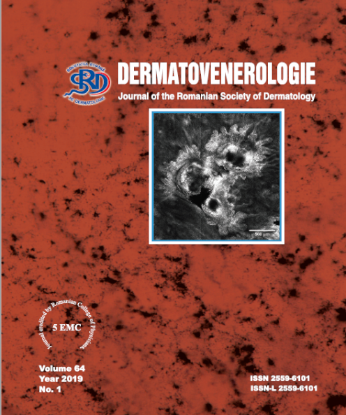
MCR este o tehnica imagistica neinvaziva ce
furnizeaza imagini ale unor sectiuni optice orizontale cu
rezolutie apropiata de cea a microscopiei optice clasice,
facilitând astfel corelatia cu dermatoscopia. Examinarea
prin MCR a unui carcinom bazocelular pigmentat ce
prezenta structuri “in spite de roata” la examenul
dermatoscopic a evidentiat prezenta de insule tumorale
hiper-refractile cu aspect floral localizate la nivelul
jonctiunii dermo-epidermice si dermului papilar, infiltrate
de celule dendritice si inconjurate de zone hipo-refractile de
clefting. Aceste insule tumorale erau conectate cu
epidermul prin multiple proiectii cu aspect de cordoane.
Rezumatin urma conferintei de consens din anul 2000 a foststabilit un set de criterii dermatoscopice semnificativepentru diagnosticul carcinomului bazocelular pigmentat(CBCp). Aceste criterii au inclus ulceratii superficiale,structuri foliacee, multipli globuli albastri-gri, cuiburi mariovoide albastre-gri, telangiectazii arborizante si structuri“in spite de roata”. Cele din urma au fost definite caproiectii radiale bine circumscrise, de culoare bruna pâna lagri, ce se intâlnesc la nivelul unui ax central de culoarebrun-inchis. Structurile “in spite de roata” au ospecificitate raportata de 100% pentru CBCp.Corelatiile morfologice intre dermatoscopie sihistologie sunt adesea anevoioase, in special datoritaincidentelor diferite din care sunt privite leziunile(orizontal in dermatoscopie vs vertical in histologie). inaceasta lucrare ne propunem sa evidentiem substratulstructurilor “in spite de roata” vizibile dermatoscopic prinutilizarea microscopiei confocale de reflectanta in vivo(MCR).MCR este o tehnica imagistica neinvaziva cefurnizeaza imagini ale unor sectiuni optice orizontale curezolutie apropiata de cea a microscopiei optice clasice,facilitând astfel corelatia cu dermatoscopia. Examinareaprin MCR a unui carcinom bazocelular pigmentat ceprezenta structuri “in spite de roata” la examenuldermatoscopic a evidentiat prezenta de insule tumoralehiper-refractile cu aspect floral localizate la niveluljonctiunii dermo-epidermice si dermului papilar, infiltratede celule dendritice si inconjurate de zone hipo-refractile declefting. Aceste insule tumorale erau conectate cuepidermul prin multiple proiectii cu aspect de cordoane.